
CT Center
All X-ray examinations done immediately on site within minutes

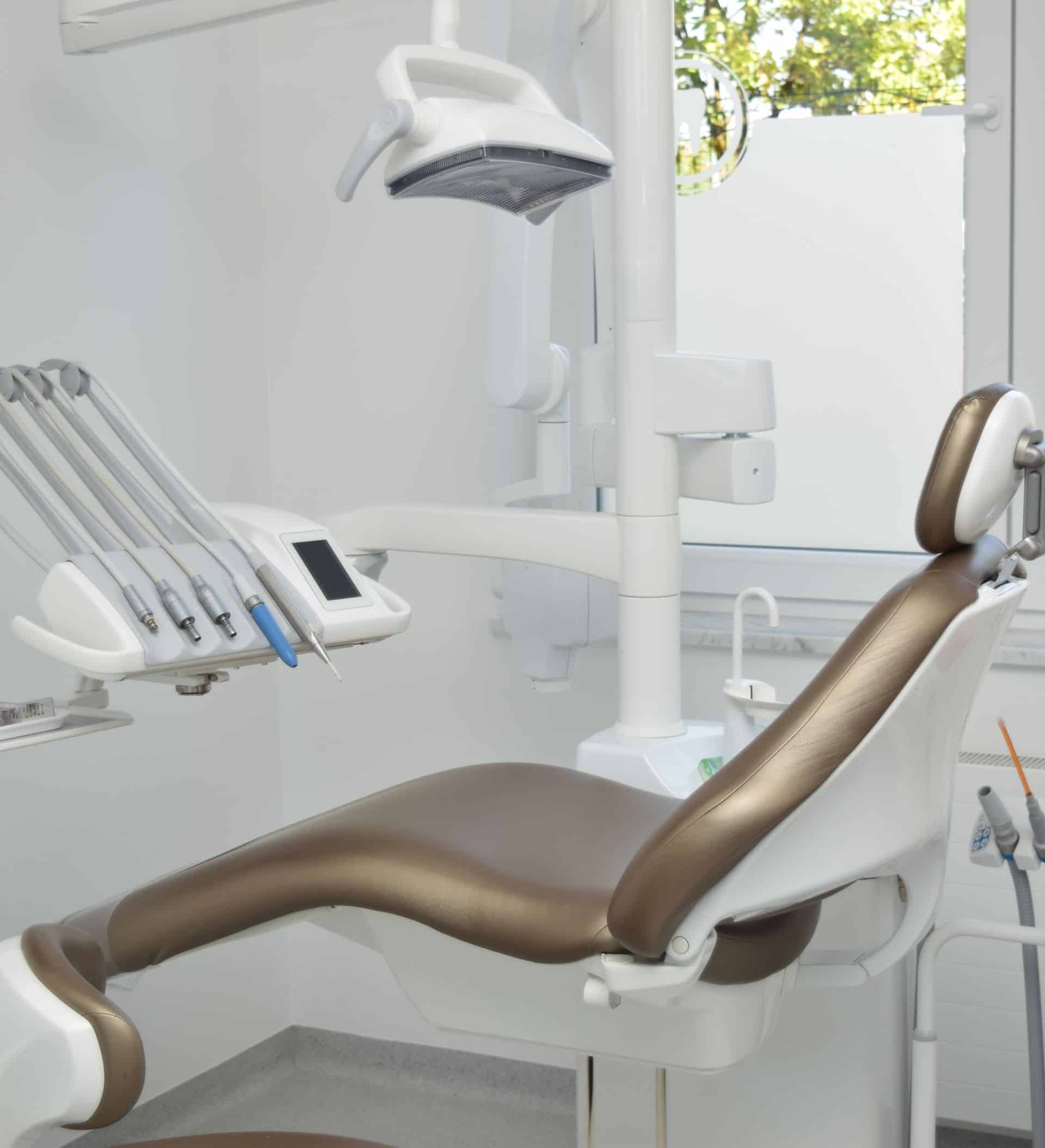
State of the art digital x-ray equipment
Do you remember the time when you first had to go to your dentist for a referral, then you had to order an appointment and get a date that generally didn’t suit you and was in your working hours, so you had to take a day off. Thanks to the modernization of X-ray devices and the availability of their procurement, these times are, thankfully, behind us all! Our dental oclinic offers you the most sophisticated digital dental x-ray, which radiates up to 90% less than the classic obsolete RTG.
In addition to the state-of-the-art equipment in the clinic, we also have our own CT center to allow patients to have all x-rays in one place without waiting in just a few minutes.
The clinic is equipped with CT 3D dental x-ray, dental orthopan and x-ray for targeted intra-oral imaging . Reading and detailed analysis are obtained minutes after the recording.
Planmeca Romexis system ProX – ProMAX3D
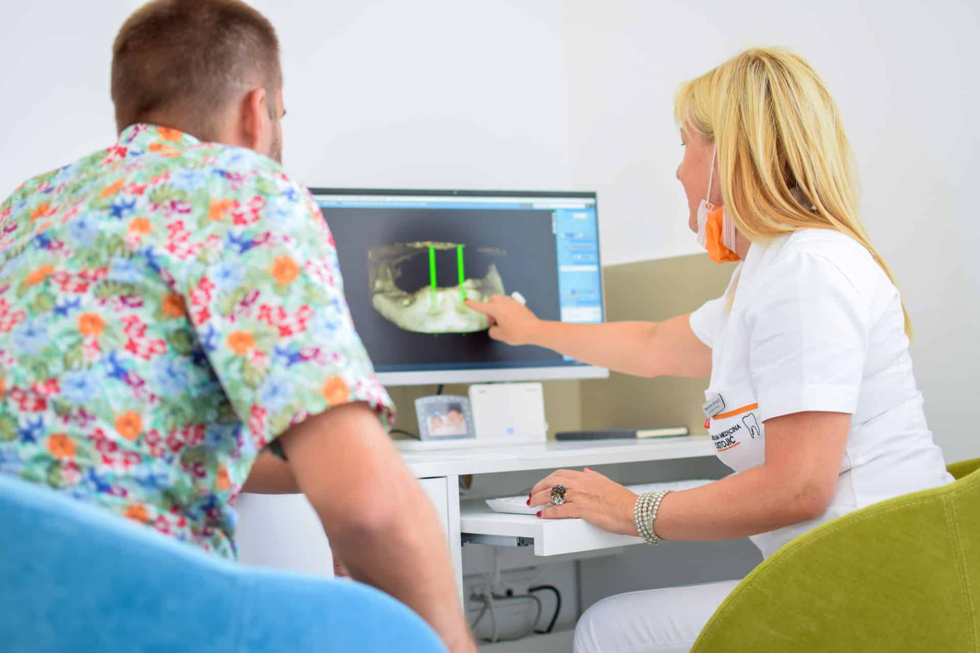
At Dental Center Ostojic, we are proud to present our modern CT center with Planmeca’s Romexis system, ProX and ProMAX3D X-ray machines. Precision, accuracy and speed adorn this system which provides detailed insight into the condition of the bone, the root of the tooth, the inter-jaw relations. Accurate diagnosis helps our dentists determine detailed treatment plans and enables patients to predict oral problems with predictability.
Digital orthopan and RVG intraoral recordings
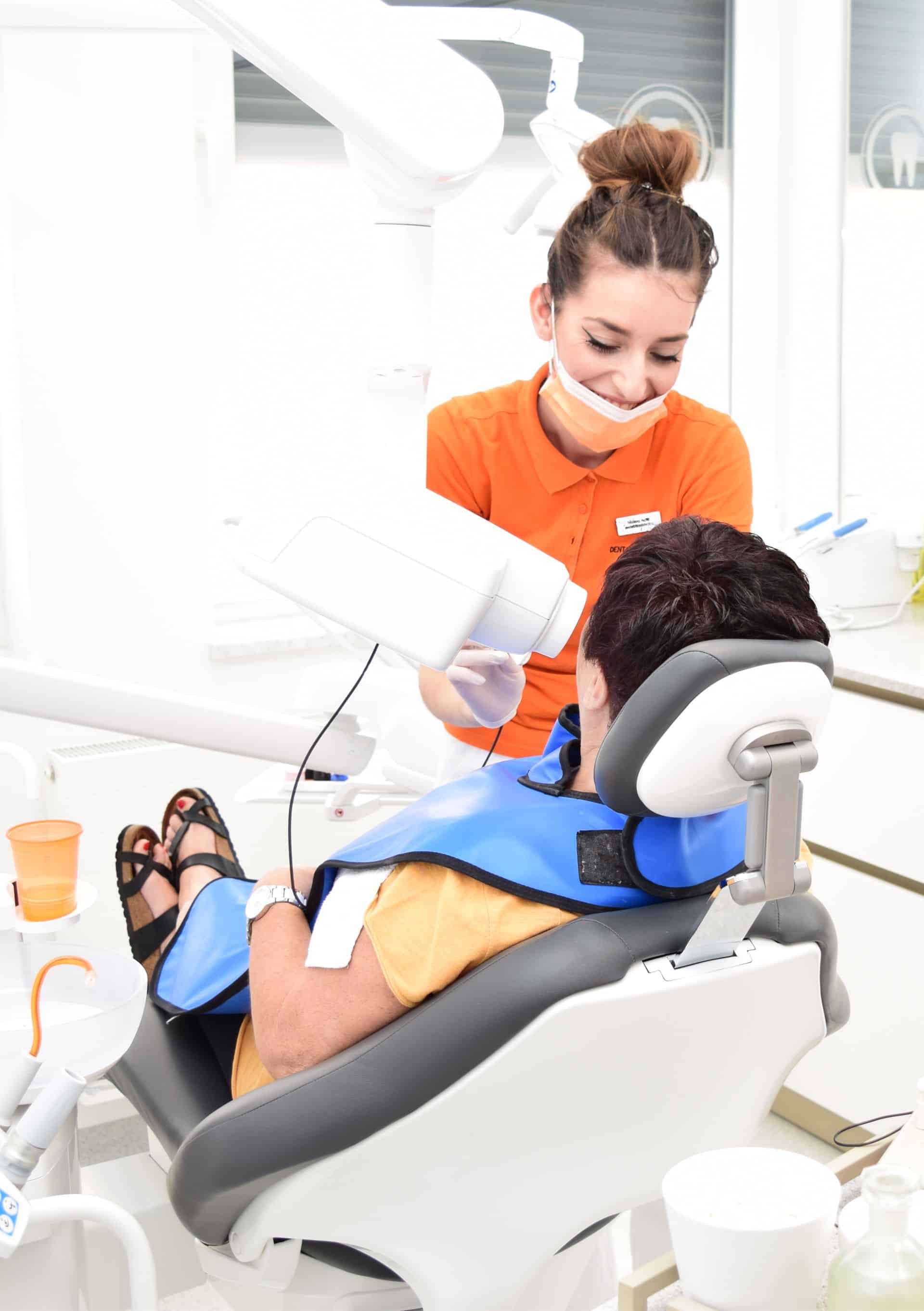
The high-precision RVG image of an individual tooth or implant provides us with detailed information about the tooth (caries progression, root canal length, accessory canals, invisible root fractures), implant, surrounding bone, and possible pathological changes.
Used when you need to get a detailed and high-quality view of a particular tooth.
RVG images are recorded using digital sensors and the amount of radiation is minimized. Recording is done on the dental chair itself, which means maximum patient comfort and extreme speed.
An orthopan is a two-dimensional X-ray of the upper and lower jaws that provides a general view of the current state of the patient’s teeth and jaws.
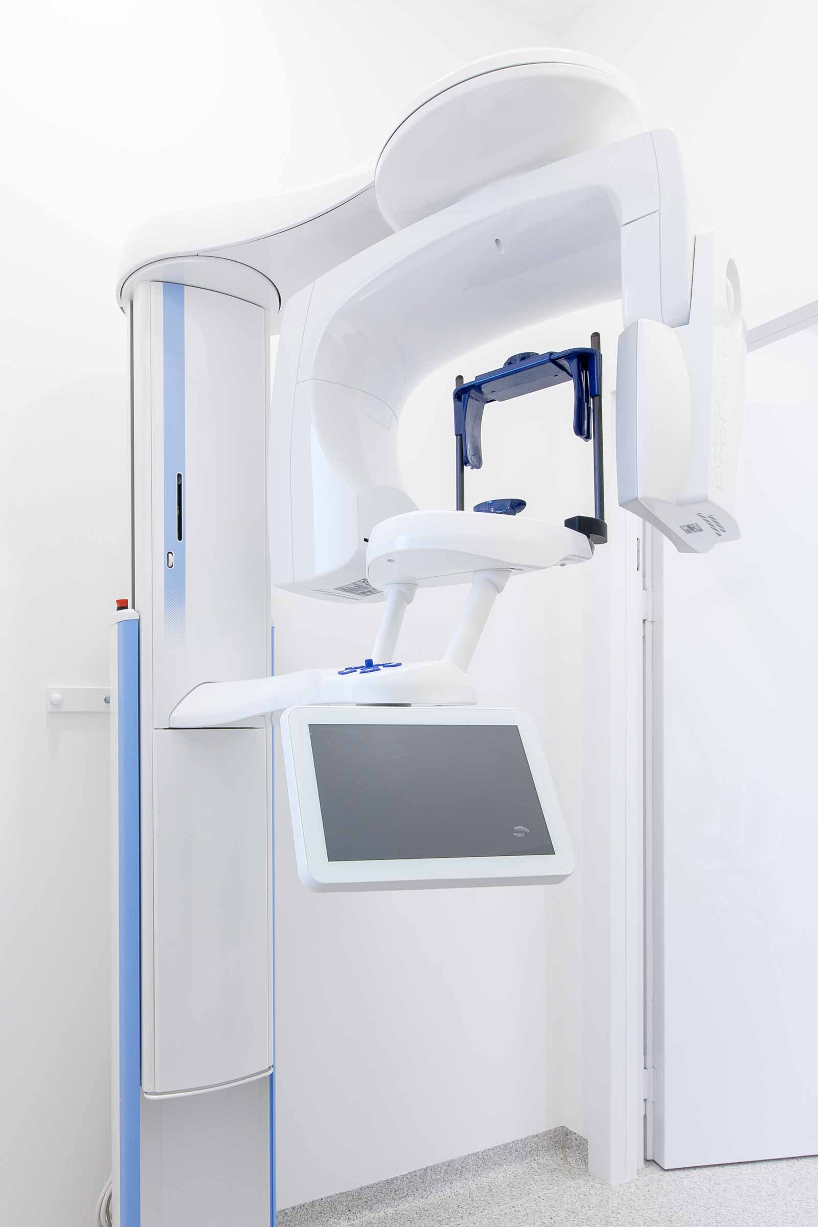
CBCT recording
Unlike classic RTG images, 3D CBCT images provide complete information on the recorded field.
In dental medicine, 3D diagnostics is provided by a Cone Beam computer tomography CT scanner.
The device displays structures with high precision with very low radiation levels. Knowledge of the third dimension of anatomical structures makes it easier to plan surgeries and make them more predictable.
It is possible to evaluate the quality (density) of the bone and familiarize each patient with the treatment plan in detail. Unlike classic RTGs, 3D imagery gives complete information about the recorded field. It is possible to effectively plan the implant procedure with respect to the bottom of the maxillary sinus, the direction and position of the mandibular canal, the width and lingual inclination of the alveolar ridge.
It is also possible to accurately diagnose various pathological formations such as periodontal pockets, cysts, root fractures, etc.…
Looking for more information?
Visit us directly at our address or contact us by email or phone
You are currently viewing a placeholder content from Google Maps. To access the actual content, click the button below. Please note that doing so will share data with third-party providers.
More InformationOur address
E-mail address
Monday - Friday
07:00 - 21:00
Saturday
Closed
Sundays and holidays
Closed
Our adress
E-mail address
Monday - Friday
07:00 - 21:00
Saturday
Closed
Sundays and holidays
Closed
You are currently viewing a placeholder content from Google Maps. To access the actual content, click the button below. Please note that doing so will share data with third-party providers.
More Information